WHEEL OF MISFORTUNE: EXTENSIVE DENTOALVEOLAR TRAUMA FOLLOWING A CYCLING ACCIDENT By Prateek Biyani I Specialty doctor in oral and maxillofacial surgery, Chesterfield Royal Hospital, Chesterfield; Daniel Shaw I Maxillofacial technician, Chesterfield Royal Hospital, Chesterfield; Alexandra Thompson I Specialty registrar in oral and maxillofacial surgery, Royal Hallamshire Hospital,Sheffield and Robert Orr I Consultant in oral and maxillofacial surgery, Chesterfield Royal Hospital, Chesterfield Leave a comment
Dentoalveolar trauma is extremely common, particularly amongst children. The majority of this trauma is managed by general dental practitioners, with few cases being referred to emergency departments.
Effective and appropriate management, following published guidelines, is important to achieve the best outcomes for patients.
We present a case report of significant dentoalveolar trauma presenting to the emergency department. We discuss immediate, short-term and long-term management, including the challenges faced along the way.
BACKGROUND
Dentoalveolar trauma is extremely common, with a prevalence of approximately 48% amongst all facial trauma.1 Attendance to emergency departments, following dental trauma, has been found to be between 4.6-10.5%.2,3
Falls are one of the most common causes of dental trauma, particularly in the younger population, closely followed by automobile and motorbike/bicycle collisions.3,4 This case report discusses an incident of significant dental trauma following a bicycle accident.
We also highlight the difficulties and challenges in immediate and short-term rehabilitation of the patient.
CASE PRESENTATION
A 13-year-old boy presented to the emergency department after going over the handlebars of his bicycle whilst not wearing a helmet. He had general symptoms of concussion but was otherwise cleared of any major head injury. He had sustained a buckle fracture of his right wrist and extensive dentoalveolar injuries. Despite the extent of trauma, he complained of minimal pain. He was fit and well, with up-to-date vaccinations and no allergies.
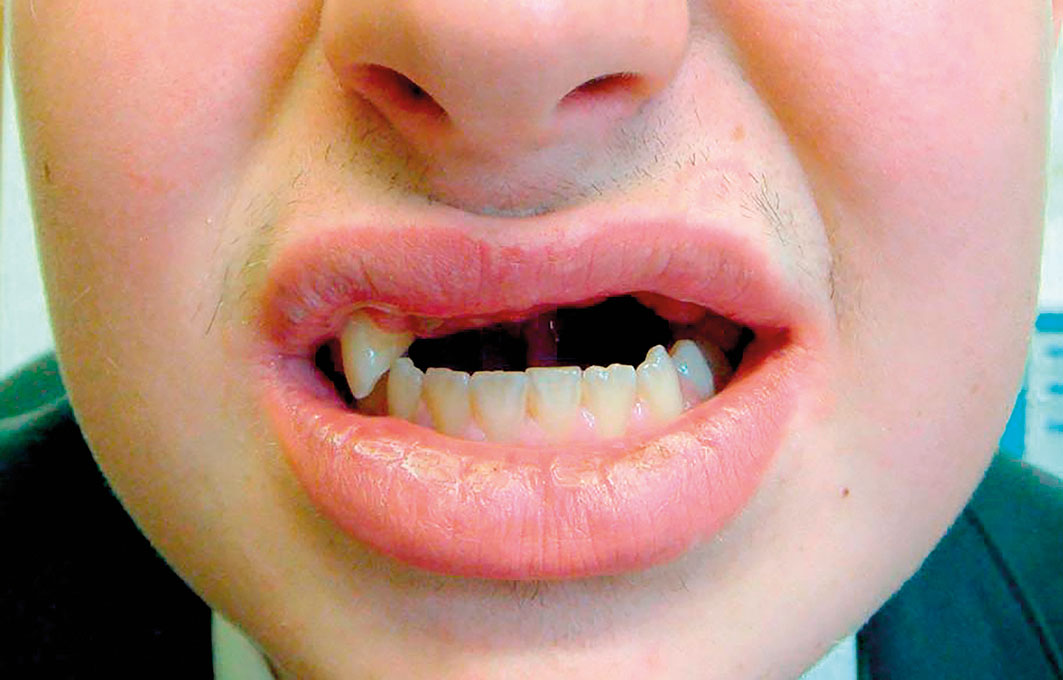
On clinical assessment of the dentoalveolar trauma, he had avulsed UR1, UL1, UL2 and UL3 along with the buccal alveolar bone and gingival tissue. This was unaccounted for and constituted a fracture of the maxillary alveolar process. Additionally, he had fractured UR2 and UL4 subgingivally (roughly to the coronal third of the root). The palatal alveolar bone and tissues were intact. Surprisingly, there were no other facial injuries of note. An orthopantomogram (OPT) was not possible on the day of presentation but a chest X-ray confirmed no aspiration of any of the avulsed tissues.
As there was very little immediate action that could be taken, local anaesthetic was given around the wound and the wound was thoroughly irrigated and debrided with saline. Sutures were not possible due to the lack of free tissue. He was prescribed a five- day course of 625 mg co-amoxiclav TDS, as well as chlorhexidine and benzydamine hydrochloride mouthwashes. He was advised to maintain a soft diet for at least four weeks and to take analgesia as required.
The patient was reviewed a week later, where an OPT was taken. He appeared to be coping well with a soft diet and the wound had begun to granulate with no signs of infection (Figure 1). The OPT corroborated the clinical findings, with the UR2 and UL4 being fractured subgingivally (Figure 2).
At this point we decided to involve the maxillofacial technician to start to consider methods of managing the wound. A cover plate was considered at this point but, as the patient was managing a sufficient oral intake and was keeping the area clean, it was decided that this would not be beneficial. Instead we decided to plan for a partial denture once more mucosal healing had occurred. The decision to wait is discussed later in the ‘Technical aspects of rehabilitation’.
We continued with weekly reviews. During this time the patient’s dentist attempted extraction of the UL4 but was unsuccessful and the tooth fractured further.
Approximately six weeks following the injury, the patient had achieved full mucosal coverage and impressions for a partial denture were commenced (Figures 3-5). He had an upper partial denture fitted with excellent aesthetics and retention (Figure 6).
Additionally, a referral was made to the joint orthodontics restorative clinic as it was assumed that the patient would require intervention from other specialities. From here, he was referred to the paediatric dentists at the Charles Clifford Dental Hospital, Sheffield, who advised to maintain the retained roots of UR2 and UL4 in situ to preserve bone. Ultimately, it was decided that he would be referred by his dentist when he turns 18 to the restorative department for dental implants.
TECHNICAL ASPECTS OF REHABILITATION
With any form of trauma, the definitive restorative device is better completed following complete healing of the hard tissues, allowing the overlying soft tissues to mature. There is need for a full clinical assessment as to the vitality of any remaining teeth that could be used as fixation structures for the final prosthesis. Furthermore, swelling of the defect area will also give a false indication of its surface topography. With these factors taken into consideration, a reasonable amount of time is given to simply allow the healing process to take place whilst the patient is under regular observation. However, these types of cases prove that treatment must be tailored to the individual and their personal requirements.
With this patient, cosmetics did not appear to be of any concern, therefore we could allow time for the area to heal fully and assess him for a prosthesis after six weeks. Had the patient wanted to wear something in the interim period then this would have been provided, however, it would have taken more appointments for adjustments/additions within the aforementioned healing process. We had considered a cover plate with attached teeth but discounted this due to the risk of causing further trauma in a patient who was functioning relatively well without a prosthesis.
This patient presented with a full permanent dentition and reasonable oral hygiene. The occlusion was well established with good interdigitation. Upon assessment it was decided to go straight to fit with a partial denture supported on a heat cured acrylic base with 0.8 mm Adams clasps on 64 I 57 (Figures 8-10). The teeth to be replaced were 21 I 1234 and these were shade matched to A2 (Dentacryl HXL).There was also greater extension of the anterior labial segment due to missing bone that was traumatised. Whilst manufacturing, consideration was given to the full extent of the labial segment and it was decided to potentially reline in Coe- Soft should extra material be required and if there was more stability deemed necessary. This would have been apparent when the patient incised whilst masticating, as the prosthesis would have tipped down at the posterior margin, compromising stability.
DISCUSSION
Dentoalveolar trauma is most common under the age of 10 and the majority (81%) of all recorded dental trauma occurs before the age of 30.1 Aetiology is variable, but by far the two most common causes are accidents (at home or while playing) and sports-related trauma (including swimming, skiing and cycling).1,3,4 Dental trauma resulting from assaults, including domestic violence, tends to peak in the second and third decades of life.1 It is important with any dentoalveolar trauma to briefly consider non-accidental injury (NAI) as 50% of physical trauma occurs in the head and neck area.1 Key dentoalveolar signs of NAI include lacerations along the buccal mucosae, a torn labial fraenum, intraoral contusions and burns on intraoral tissues.5 Sports-related dentoalveolar trauma is common in the community, accounting for 50% of all sports-related facial injuries.1 One study found that 13.6% of dentoalveolar trauma that passed through their emergency department specifically cited bicycle-related trauma, much like our presented case.4
The two most common forms of dentoalveolar trauma appear to be crown fractures and subluxations.1,2 Fractures of the alveolar process are relatively rare with various studies reporting a prevalence of 2-9.8%.2,3,6 This particular case would constitute a segmental fracture, which results from more severe trauma and involves multiple teeth and their supporting alveolar process.5 The majority of cases with a fractured alveolar process impact the maxilla, and typically involve just two teeth.6
Our case involved avulsion of four teeth and extensive fractures of two, which is extremely rare. Avulsion, along with the alveolar fracture, has been noted in around 11% of cases.6 The rarity of such severe dentoalveolar trauma poses great challenges and complexity in management.
Management of this patient had to be considered in three phases – immediate term, short-term and long-term. Like with any form of dentoalveolar trauma, our first consideration was the Dental Trauma Guidelines set out by the International Association of Dental Trauma (IADT).7 According to these guidelines, the ideal management for this case (had the entire segment not avulsed) would have been to reposition the segment and splint it for four weeks. Along with this, basic conservative advice of a soft diet, analgesia, antibiotics and good oral hygiene should be stressed. Following this, close clinical follow-up would be necessary. Due to the lack of segment when this patient attended, we were only able to intervene with basic clinical measures and debride the wound. The lack of loose soft tissue prevented us from placing any sutures.
When considering short and long-term management for this patient, the priorities are function and aesthetics. Dentoalveolar trauma has been found to have major impacts on children’s quality of life (QoL), comparable to patients with cleft lip and palate.8,9 Functional limitations, including taking longer to eat and difficulty with speech, have been reported as being of significant concern to dentoalveolar trauma patients.9 Surprisingly, aesthetics were not a concern for our patient. However, generally it has been found that aesthetics is of a greater concern to children than function.10 This also has implications on the emotional wellbeing of children. A large proportion of younger children were found to be upset and shy following their trauma, whilst older children exhibited frustration and concern about how others would see them.9
In order to rehabilitate this patient, in terms of both aesthetics and function, we decided to provide the patient with a removable partial denture. This provided multiple advantages including the ability to reline and adjust the denture during healing, as well as utilise it for treatment planning in the future. However, removable partial dentures are a compromise at this age and ideal rehabilitation would be with dental implants and implant supported restorations. Due to the age of the patient, definitive rehabilitation was not feasible as he was in a growth phase. Children who have had removable partial dentures tended to have more complaints in relation to eating and social interaction.11 For long- term rehabilitation with dental implants, most children will require some type of bone augmentation procedure, such as a bone graft.12 This is something we made the patient aware of early on. This patient was referred to the orthodontist to determine whether orthodontic extrusion of the remaining fractured teeth may preserve some bone. Ultimately it was determined that there would be no major benefit of extruding the UR2 and UL4, and instead, maintaining them in situ for as long as possible would be sufficient. Success rates of anterior maxillary implants placed with bone grafting have been found to be around 96.8%, suggesting that this would be a predictable long-term solution for these patients.13
Management of dentoalveolar trauma is challenging in most situations. The extent and severity of trauma will heavily influence treatment planning and decision-making. Dentoalveolar trauma can have impacts physically, mentally and on quality of life. As demonstrated in this case, compromises may sometimes need to be made in the short-term in order to achieve function and aesthetics. Ultimately, the use of implant supported restorations will provide the best and most predictable results for these patients.
DECLARATION OF INTERESTS
We have no conflicts of interest in this case report.
REFERENCES
- Gassner R, Bösch R, Tuli T, Emshoff R (1999). Prevalence of dental trauma in 6000 patients with facial injuries. Oral Surgery, Oral Medicine, Oral Pathology, Oral Radiology, and Endodontology, 87(1): 27-33.
- Galea H (1984). An investigation of dental injuries in an acute care general hospital. The Journal of the American Dental Association, 109(3): 434-438.
- Luz J, Mase F (1994). Incidence of dentoalveolar injuries in hospital emergency room patients. Dental Traumatology, 10(4): 188-190.
- Blinkhorn F (2000). The aetiology of dento-alveolar injuries and factors influencing attendance for emergency care of adolescents in the North West of England. Dental Traumatology, 16(4): 162-165.
- Dale R (2000). Dentoalveolar Trauma. Emergency Medicine Clinics of North America, 18(3): 521-538.
- Andreasen J, Lauridsen E (2015). Alveolar process fractures in the permanent dentition. Part 1. Etiology and clinical characteristics. A retrospective analysis of 299 cases involving 815 teeth. Dental Traumatology, 31(6): 442-447.
- Andersson L (2012). IADT guidelines for treatment of traumatic dental injuries. Dental Traumatology, 28(1): 1-1.
- Traebert J, de Lacerda J, Foster Page L, Thomson W, Bortoluzzi M (2012). Impact of traumatic dental injuries on the quality of life of schoolchildren. Dental Traumatology, 28(6): 423-428.
- Berger T, Kenny D, Casas M, Barrett E, Lawrence H (2009). Effects of severe dentoalveolar trauma on the quality of life of children and parents. Dental Traumatology, 25(5): 462-469.
- Ilma de Souza Cortes M, Marcenes W, Sheiham A (2002). Impact of traumatic injuries to the permanent teeth on the oral health-related quality of life in 12-14- year-old children. Community Dentistry and Oral Epidemiology, 30(3): 193-198.
- Bouchardet F, Ilma de Souza Gruppioni Cortes M, Vilela Bastos J, Alexandra Costa de Morais Caldas I, Franco A, Nuno Pessoa Vieira D (2014). The impact of tooth avulsion on daily life performance using the Brazilian OIDP index in children and young adults. Journal of Forensic Odontostomatology, 32(1): 9-14.
- Schwartz-Arad D, Levin L (2004). Post-traumatic use of dental implants to rehabilitate anterior maxillary teeth. Dental Traumatology, 20(6): 344-347.
- Nissan J, Gross O, Mardinger O, Ghelfan O, Sacco R, Chaushu G (2011). Post-traumatic implant-supported restoration of the anterior maxillary teeth using cancellous bone block allografts. Journal of Oral and Maxillofacial Surgery, 69(12): e513-e518.